-
Monera , Protista , Fungi
KINGDOM - MONERA
Christesen & dougherty - gave the term ‘prokaryote’ and ‘eukaryote’
C.B. Van Neil : Divided the living organisms into prokaryotes and eukaryotes.
Main characteristic of prokaryotes :
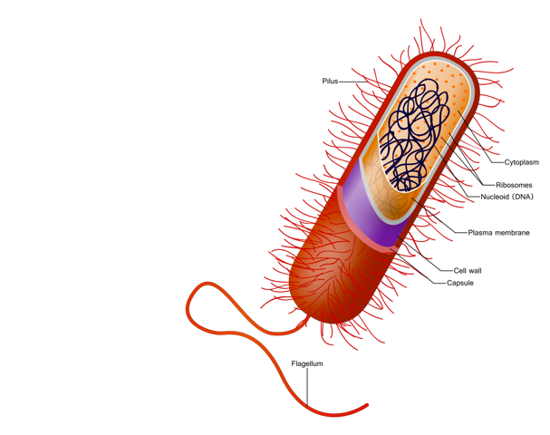
(1) Cell wall :
Cell wall of prokaryotes is made up of peptidoglycan (or murein) which is a type of mucopeptide.
The structure of peptidoglycan has two parts
(A) Polysaccharides : They are made up of glucose, mannose, galacotse and amino sugar units. Amino sugars → N-acetlyl glucosamine, N-acetyl muramic acid
(B) Amino acids : Following amino acids are present in the cell wall of prokaryotes : (i)L - alanine (ii) L - Lysine (iii) D- alanine (iv) D- glutamic acid
1. Diaminopimelic acid - This non proteineous amino acid is found in all eubacteria (Gram + & Gram -) and blue green algae.
2. Muramic acid - This acid is found in eubacteria and BGA
3. Teichoic acid - This acid is found only in Gram (+) eubacteria.
Note : The structure of cell wall in prokaryotes is complicated and this is the primitive character.
Generally the cell wall of prokaryotes is not made up of cellulose but exceptionally there are some prokaryotes in which cell wall made up cellulose, which are as follows -
eg. Acetobacer acetogenum Acetobacter Xylenum Zymosarcina
Prokayotes which are intra cellular lack cell wall
eg. Mycoplasma
(2) Cell membrane :
(A) Like eukaryotes the cell membrane of prokaryotes is made up of lipoprotein [lipid + protein]
(B) The space between cell wall and cell membrane is known as periplasmic space. This space, is analogus to lysomome because in this space the digestion of complex substance is done.
(3) Cytoplasm :
(A) The cytoplasm of prokaryotes lacks membrane bound cell organelles.
(B) In Prokaryotic cell, the nucleus is indistinct. The nucleus of prokaryotes is also known as incipient nucleus, genophore, nucleiod or fibrillar nucleus. Nuclear membrane is absent around nucleus. It also lacks nucleolus.Prokaryotes also lack the true chromosome. Instead of it, a false chromosome is present, which is made up of circular naked DNA + Non histone protein. Non histone proteins are polyamines. This false chromosome coils and forms the chromosomal region, which is known as nucleoid.
(C) Is prokaryotes ribosomes are of 70s type.
(4) Movement :
prokaryotes are both motile & Non motile
(A) Motile prokaryotes -
In prokaryotes two types of locomotion are present
Gliding : Locomotion by slipping → They secrete mucilage and then glide on this mucilage. There is no particular structure like cilia, flagella for locomotion.
eg. Myxobacteria, Cyanobacteria (BGA)
Swimming : Flagella are present in many prokaryotes for swimming.
eg. Eubacteria
(B) Non motile prokaryotes -
eg. Mycoplasma
EUBACTERIA
They were first observed in rainy water and later in teeth scum by Leeuwenhoek (1675) and called them “Animalcule”. This discovery was published in his book “The Secrets of nature Discovered by Leeuwenhoek”
F.H. Cohn and Ehrenberg first of all coined the name “Bacteria”.
Bergey placed bacteria in “Prosophyta group’ and wrote a book “Munual of Determinative Microbiology”. This book is known as “Bible of bacterial classification”.
Lister developed “culture technique”. He also developed the “sterilization tech” to sterlize the surgical instruments. He discovered the antiseptic nature of carbolic acid. Lister first of all cultured bacteria artificially.
Louis Pasteur proposed “germ theory” and called the bacteria “germ”. He discovered the “Pasteurisation technique”. (Pasteurisation technique - it is a process which means heating of drinks. it is carried out at 600C temperature and for 30 minutes duration).
Robert Koch
Koch first obtained pure culture of bacteria.
He discovered the Anthrax. T.B. and Cholera causing bacteria.
Koch gave some rules to prove that the bacteria are the cause of disease. These rules are known as “Koch postulates”.
He awarded “Nobel Prize” for his work.
Koch postulates do not applicable on obligate parasite (eg. Mycobacterium leprae)
SIZE
Smallest eubacteria = Haemophilus influenzae → 0.2 - 0.3 × 0.5 - 2.0 micrometer
Longest / largest eubacteria = Epulopiscium fishelsoni → 600 micrometer
Largest / longest Filamentous bacterium = Beggiatoa mirabilis → few mm.
SHAPE
Bacteria have variation in their shape. On the basis of their shape bacteria are of different types.
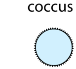
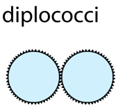
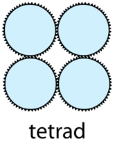
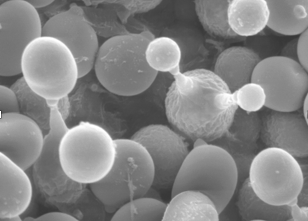
1. Coccus (Pl. Cocci → Sing. Coccus)-
These bacteria are spherical
These are smallest bacteria
These are highly (Maximum) resistant.
These are following types
A. Monocossus - These spherical bacteria live alone (single sphere) e.g. Micrococcus, Dialister pneumosintes
B. Diplococcus - These are found in pair. e.g. Diplococcus pneumniae, Neisseria
C. Tetra occurs - These are found in group of four cocci. e.g. Micrococcus luteus
D. Streptococcus - These are found in form of chain . e.g. Streptococcus lactis
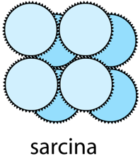
E. Sarcinae - 8 to 64 or ore bacteria are found in cubical mass form . e.g. Sarcina
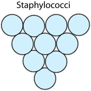
F. Staphylococcus - These bacteria are found in a irregular bunch . e.g. Staphylococcus alvus

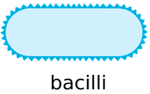
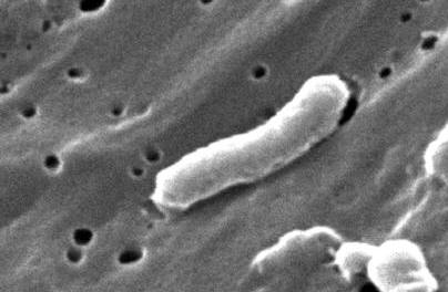
2. Bacillus (Pl. Bacilli - Sing. Bacillus)-
This group includes most of the bacteria.
These are rod shaped
They are following types
A. Single Bacillus - Only one rod -like structure or bacterium. e.g. E.coli, Lactobacillus
B. Diplobacillus - They are found in pairs e.g. Diplobacillus

C. Streptobacillus - They are found in a chain e.g. Bacillus anthracis
Bacillus subtilis -It is surrounded by mucilagenous sheath that is known as zooglea. It is also known as hay bacteria.

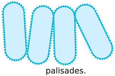
D. Palisade bacillus - These roed shaped bacteria are found in form of stacks e.g. Corynebacterium diphtheriae
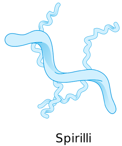
3. Spirillum (Pl. Spirilli - Sing. Spirillum) : These are spiral shaped bacteria e.g. Spirillum volutans, Spirochete, Helibacter, Treponema
4. Comma (Vibrio) :These are comma shaped bacteria. e.g. Vibrio cholerae, Vibrio comma
5. Stalked bacteria - These are single called bacteria with narrow stalked e.g. Caulobacter
6. Budding bacteria - They appear like a beaded cell e.g. Rhodomicrobium
7. Pleomorphic bacteria - These bacteria change their shape according to the medium. e.g. Rhizobium
Rhizobium bacterium is found in three forms X,T and Z
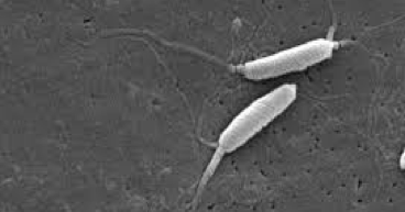
MOTILITY IN BACTERIA
Bacteria are motile as well as non motile. Movement in bacteria takes place by means of flagella.
On the basis of flagella bacteria are of following types
1. Atrichous - When flagella are absent, it is called atrocious form e.g. Micrococcus, Pasteurella
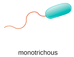
2. Monotrichous - When only one flagellum on one end of the bacterium e.g. Vibrio, Thiobacillu, Pseudomonas
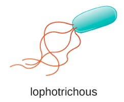
3. Lophotrichous - When a bunch of flagellum is present on one end of bacterium. e.g. Salmonella
4. Amphitrichous - When bunch of flagellum or single flagellum are present on both the ends of bacterium. e.g. Spirillu, Nitrosomonas

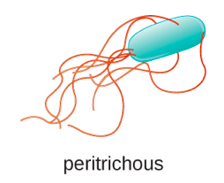
5. Peritichous - When flagella are found on the whole body of bacterium . e.g. E. coli. Salmonella typhi
NUTRITION
Most of the bacteria are heterotrophic but some are autotrophic. On the basis of nutrition bacteria are classified into following three categories.
Autotrophs
These bacteria use light or chemical energy for their own food synthesis
(i) Photosynthetic autotrophs - These bacteria use light energy for food synthesis.
e.g. Chromatium , Chlorobium, Thiothrix , Rhodospirillu, Rhodopseudomonas , Blue green algae
(ii) Chemosynthetic autotrophs -
These are nonphotosynthetic autotrophs i.e., photosynthetic pigments are absent.
They use chemical energy instead of light energy for food synthesis.
Iron bacteria - These bacteria convert Ferrous compounds into Ferric compounds . Fe+2 → Fe+3 + energy
e.g. Ferrobacillu, Leptothrix
Sulphur bacteria - convert the H2S into Sulphur and water. 2H2S + O2 → 2S + 2H2O + energy
e.g., Thiobacillus, Beggiatoa
Carbon bacteria - Convert CO into CO2 2CO + O2 → 2CO2 + energy
e.g. Bacillus oligocrbophylus
Nitryfying bacteria - They oxides nitrogenous compounds and obtain energy
e.g. Nitrosomonas , Nitrobacter
Hydrogen bacteria - Convert the hydrogen into water 4H2 + CO2 → CH4 + 2H2O + energy
e.g. Bacillus pentotrophus, Hydrogenomonas
(b) Chemo - organotrophs - These bacteria oxidise the organic compounds and released energy is used for food synthesis
e.g. methanomonasHeterotrophs
Most of the bacteria are heterotrophic i.e., they can not manufacture their own food
They receive their own food from dead organic matter or living organism
(i) Saprotrophic bacteria - These bacteria obtain food from dead and decarying organic matter
e.g. Bacillus vulgaris , Pseudomonas
(ii) Parasitic bacteria - They obtain their food from living organism
e.g. Mycobacterium leprae , Mycobacterium tuberculosis
Symbiotic bacteria
These bacteria convert atmospheric nitrogen into nitrogenous compounds like Amino acid NO3 or Salts of ammonia
e.g. Rhzobium
REPRODUCTION
A) Vegetative reproduction
(i) By budding : e.g., Hyphomicrobium vulgare, Rhodomicrobium vannielia, etc.
(ii) Binary fission : e.g., Salmonella , E.coli etc
B) Asexual reproduction
(i) By Endospore - e.g. Endospore formation is seen in mostly Bacillus type of bacteria
(ii) By Cyst - e.g., many members of Azotobacter
C) Genetic recombination
(i) Transformation - e.g., Diplococcus pneumoniea
(ii) Conjugation - e.g., E.coli
(iii) Sexduction – e.g., Salmonella typhimurium
ARCHAEBACTERIA
Evolutionary they are primitive. They were the first to be born on our planet and they are present nowdays with their primitive characters. They are the “Oldest living fossils”.
All archaebacteria are bligate anaerobes.
Thermococcus, methanococcus and methanobacteriaum exemplify archaebacteria that contain protein homologous to eukryotic core histones.
Their cell wall is not made up of peptidoglycan like that of eubacteria. Their cell wall is made up of complex polysacchrides and complex polypeptide.
Their cell membrane is not a unit membrane, while in eubacteria the cell membrane is unit membrane.
In archaebacteria sequence of nucleotide in 16 s - r RNA is differ from other prokaryote
Archaebacteria includes following bacteria
1. Methanogens : “Methane producing bacteria”
eg. Methanobaterium, methanococcus, methanomicrobium.
2. Halophiles : These archaebacteria are found in highly saline areas.
eg. Halobacterium , Halococcus
3. Thermo acidophiles : These archaebacteria are found at those places where temperature is approx 800C and medium is cidic [pH = 2]
eg. Thermus, Sulpholobus, Thermoplasma
KINGDOM - PROTISTA
Protista (Protistos = Primary) includes unicelluar eukaryotes and show the following characters :
Protists include solitary unicellular or colonial unicellular eukaryotic organisms which not form tissues.
Simple multinucleate organisms or stages of life cycles occur in a number of groups.
The organisms possess nuclear membranes and mitochondria.
In many forms, plastids, (9+2 strand) flagella and other organelles are present.
The nutritive modes of these organisms include photosynthesis, absorption, ingestion and combination of these.
Some protists possess contractile vacuole for regulation of their water content.
Their reproductive cycles typically include both asexual divisions of haploid forms and true sexual processes with karyogamy and meiosis.
The organisms move by flagella or by other means or are non-motile.
Nutrition : It is may be photosynthetic, holotrophic, saprotrophic and parasitic. Some have mixotrophic nutrition (holotrophic + saprobic). Chemosynthetic nutrition is lacking. Certain protozoans decompose organic matter, such as cellulose, in the gut of termites and woodroaches. They live as symbionts. The photosynthetic, floating protists are collectively called phytoplankton. They usually have a cell wall. The free-floating, holozoic protozoans are collectively termed zooplankton. They lack cell wall to allow ingestion of particulate food.
Reproduction : It is occurs by both asexual and sexual methods :
(i) Asexual reproduction : It is the most common method of reproduction in protists in which the genetic constitutions of young ones remains the same as that of the parent. Under favourable environmental conditions, they reproduce asexually several times a day resulting in population explosions. The major types of asexual reproductions are as follows :
(a) Binary fission : The parent cell divides into two approximately equal daughter cells either transversely (e.g., Paramecium), longitudinally (e.g., Euglena) or axially (e.g., Amoeba) by mitosis.
(b) Multiple fission : Division of parent cell into a number of daughter cells is called multiple fission. It occurs in Amoeba.
(c) Plasmotomy : Fission of multinucleate protist into two or more multinucleate offsprings by the division of cytoplasm without nuclear division is called plasmotomy. It occurs in Opalina.
(d) Budding : In this type of asexual reproduction, a small bud is formed from the parent body which separates and develops into new individual. e.g., Paracineta, Arcella, etc.
(e) Spore formation : Sessile or stalked sporangia containing spores are formed in slime moulds. They liberate the spores which can withstand a prolonged period of desiccation. On germination, each spore gives rise to new individual. e.g., Slime moulds.
(ii) Sexual reproduction : Sexual reproduction is believed to have originated in primitive protists. It involve meiosis (reduction division) and syngamy. It occurs following types.
(a) Isogamy : The two fusing gametes are structurally and functionally similar, e.g., Monocystis.
(b) Anisogamy : The two fusing gametes are similar but differ only in their size and/or motility, e.g., Ceratium.
(c) Oogamy : Large non-motile gametes are fertilized by smaller motile gametes, e.g., Plasmodium.
Major group of protists : Unicellular protists have been broadly divided is to three major grous
(i) Photosynthetic protists : Protistan algae e.g. Dinoflagellates (i.e. Ceratium, Glenodinium, Gymnodinium, Gonyaulax, Noctiluca and Peridinium), Diatoms (Navicula, Nitzchia, Melosira, Cymbella, Amphipleura, Pinnularia) and Euglenoids or Euglena like flagellates (Euglena, Eutreptia, Phacus, Peranema).
(ii) Consumer protists : Slime moulds or Myxomycetes, e.g., Physarum, Physarella.
(iii) Protozoan protists : It is include four phyla – Zooflagellata (e.g., Trypanosoma, Giardia, Trichonympha, Trichomonas, Leishmania etc.), Sarcodina (e.g., Amoeba, Entamoeba, Pelomyxa, Mestigamoeba etc.), Sporozoa (e.g., Plasmodium, Monocystis, Eimeria etc. all are endoparasites) and Ciliata (e.g., Paramecium, Vorticella, Opalina, Podophyra etc.).
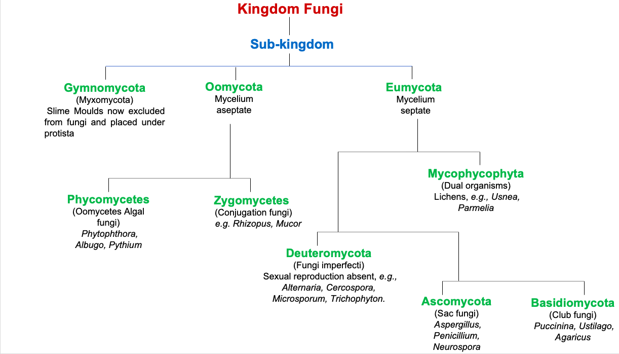
KINGDOM - FUNGI
Introduction :
The science dealing with the study of fungi is called as mycology. The knowledge of fungi to mankind dates back to prehistoric times. Clausius, 1601 may be regarded as one of the earliest writers to describe fungi. Bauhin (1623) also included the account of known fungal forms in his book Pinax Theatric Botanica. The fast systematic account of fungi came from Pier Antonio Micheli (1729) who wrote 'Nova Plantarum Genera'. He is described by some workers as founder or mycology. Linnaeus (1753) also included fungi included fungi in his 'Species Plantarum'. Elias Fries (1821-31) gave a more detailed account of fungi in his 'Silloge Fungorum' in 25 volumes describing some 80,000 species of fungi. This work remains unparalleld even today.
Thallus organization :
The plant body of true fungi (Eumycota), the plant body is a thallus. It may be non-mycelial or mycelial. The non-mycelial forms are unicellular, however, they may form a pseudomycelium by budding. In mycelial forms, the plant body is made up of thread like structures called hyphae (sing. hypha). The mycelium may be aseptate (non-septate) or septate. When non-septate and multinucleate, the mycelium is described as coenocytic. In lower fungi the mycelium is non-septate e.g., Phycomycetae. In higher forms it is septate e.g., Ascomycotina, Basidiomycotina and Deuteromycotina. In some forms the plant body is unicelled at one stage and mycelial at the other. Their organization is sometimes described as dimorphic.
Cell organization :
The cell wall of fungi is mainly made up of chitin and cellulose. While chitin is a polymer of N-acetyl glucosamine, the celulose is polymer of d-glucose. Precisely, the cell wall may be made up of cellulose-glucan (Oomycetes), chitin chitosan (Zygomycetes) mannan-glucan (Ascomycotina), chitin-mannan (Basidiomycotina) or chitin-glucan (some Ascomycotina, Basidiomycotina and Deuteromycotina). Besides, the cell wall may be made up of cellulose-glycogen, cellulose-chitin or polygalactosamine-galactan.
Nutrition :
The fungi are achlorophyllous organisms and hence they can not prepare their food. They live as heterotrophs i.e., as parasites and saprophytes. Some forms live symbiotically with other green forms.
(i) Parasites : They obtain their food from a living host. A parasite may be obligate or facultative
(ii) Saprophytes : They derive their food from dead and decaying organic matter. The saprophytes may be obligate or facultative
(iii) Symbionts : Some fungal forms grow in symbiotic association with the green or blue-green algae and constitute the lichen. Here the algal component is photosynthetic and the fungal is reproductive. A few fungal forms grow in association with the roots of higher plants. This association is called as mycorrhiza.
Reproduction : The fungi may reproduce vegetatively, asexually as well as sexually
(i) Vegetative reproduction
(a) Fragmentation : Some forms belonging to Ascomycotina and Basidiomycotina multiply by breakage of the mycelium.
(b) Budding : Some unicelled forms multiply by budding. A bud arises as a papilla on the parent cell and then after its enlargement separates into a completely independent entity.
(c) Fission : A few unicelled forms like yeasts and slime molds multiply by this process.
(d) Oidia : In some mycelial forms the thallus breaks into its component cells. Each cell then rounds up into a structure called oidium (pl. oidia). They may germinate immediately to form the new mycelium.
(e) Chlamydospores : Some fungi produce chlamydospores which are thick walled cells. They are intercalary in position. They are capable of forming a new plant on approach of favourable conditions.
(ii) Asexual reproduction
(a) Sporangiospores : These are thin-walled, non-motile spores formed in a sporangium. They may be uni-or multinucleate. On account of their structure, they are also called as aplanospores.
(b) Zoospores : They are thin-walled, motile spores formed in a zoosporangium. The zoospores are of several types :
Uniflagellate with whiplash type flagellum e.g., Allomyces.
Uniflagellate with tinsel type flagellum e.g., Rhizidiomyces.
Biflagellate with a tinsel type and a whiplash type flagella e.g., Saprolegnia.
Biflagellate with two whiplash type flagella e.g., Plasmodiophora.
(c) Conidia : In some fungi the spores are not formed inside a sporangium. They are born freely on the tips of special branches called conidiophores. The spores thus formed are called as conidia. On the basis of development, two types of conidia are recognised namely thallospores and blastospores or true conidia.
Thallospores : In some forms the thallus itself forms spore like bodies called thallospores. The thallospore are of two types namely arthrospores and chlamydospores.
Arthrospores : They are thinwalled spores formed in basipetal order e.g., Endomyces.
Chlamydospores : Some of the hyphal cells are converted into thick walled chlamydospores. They may be terminal or intercalary e.g., Ustilago, Saprolegnia.
Blastospores : They develop on conidiophores in acropetal or basipetal succession. They are of two types –
Porospores : When the blastospores develop by the balooning of the inner wall of conidiophore, it is called as porospore e.g., Alternaria.
Phialospore : On the other hand, when the first conidium carries the broken parent wall of conidiophore and subsequent conidia possess a new wall, such basipetally formed conidia are called as phialospore e.g., Aspergillus.
(iii) Sexual reproduction : With the exception of Deuteromycotina (Fungi imperfect), the sexual reproduction is found in all groups of fungi. During sexual reproduction the compatible nuclei show a specific behaviour which is responsible for the onset of three distinct mycelial phases. The three phases of nuclear behaviour are as under :
Plasmogamy : Fusion of two protoplasts.
Karyogamy : Fusion of two nuclei.
Meiosis : The reduction division.
Salient features of classes
(i) Phycomycetes (Oomycetes/Egg fungi) :
It is also called lower fungi, mycelium is coenocytic. Hyphal wall may contain chitin or cellulose (e.g., Phytophthora). Asexual reproduction occurs with the help of conidio-sporangia. Under wet conditions they produce zoospores. Under dry conditions, the sporangia directly function as conidia. Zoospores have heterokont flagellation (one smooth, other tinsel). Sexual reproduction is oogamous. It occurs by gametangial contact where male nucleus enters the oogonium through a conjugation tube. The fertilized oogonium forms oospore. e.g., Sapolegnia, Albugo (Cystopus), Phytophthora, Phythium, Sclerospora.
(ii) Zygomycetes (Conjugation fungi) : Mycelium is coenocytic. Hyphal wall contains chitin or fungal cellulose. Motile stage is absent. Spores (Sporangiospores/aplanospores) are born inside sporangia. Sexual reproduction involve fusion of coenogametes through conjugation (Gametangial copulation). It produces a resting diploid Zygospore. On germination, each zygospore forms a germ sporangium at the tip of a hypha called promycelium e.g., Mucor, Rhizopus.
(iii) Ascomycetes (Ascus : sac, mycete : fungus) : These are unicellular as well as multicellular fungi. In the latter, mycelium is septate. The asexual spores formed in chains are called conidia. The spores are formed exogenously, i.e. outside sporangium. They detach from the parent and form new mycelia. Sexual reproduction is through ascospores, which are formed endogenously (within the mycelium) in a sac like structure called ascus (pl. asci). The gametes involved in sexual reproduction are nonmotile compatible and are generally represented as + and –. The fusion of gametes is followed by reductional division that produces haploid ascospores. The fruiting body called ascocarp.
The ascocarp are of four types :
(a) Cleistothecium : It is an ovoid or spherical fruiting body which remains completely closed e.g., Aspergillus.
(b) Perithecium : It is a flask shaped fruiting body which opens by a single pore called ostiole. It is lined by sterile hyphae called paraphyses. The asci are also mixed with paraphysis e.g., Cleviceps.
(c) Apothecium : It is a saucer-shaped fruiting body. The asci constitute the fertile zone called hymenium e.g., Peziza.
(d) Ascostroma : It is not a distinct fruiting body. It lacks its own well defined wall. The asci arise directly with a cavity (locule) of stroma. It is also called as pseudothecium e.g., Mycosphaerella.
(iv) Basidiomycetes : They are the most advanced fungi and best decomposers of wood. These are called club fungi because of a club shaped end of mycelium known as basidium. They have septate multinucleated mycelium. Septa possess central dolipores and Lateral clamp connections. The sexual spores called basidiospores are generally four in number. They are produced outside the body (exogenuous) unlike ascomycetes where they are endogenous. Two compatible nuclei fuse to form zygote, which undergoes meiosis and forms four basidiospores. The fruiting body containing basidia is a multicelular structure called basidiocarp. The common members are edible mushrooms (Agaricus), Smut and Rust.
(v) Deuteromycetes (Fungi inperfecti) : The group include all those fungi in which sexual or perfect stage is not known. Mycelium is made of septate hyphae. Asexual reproduction commonly occur by means of conidia.